Quick Facts
- Category: Science & Space
- Published: 2026-05-04 21:33:25
- Linux Kernel Patch Promises Better Gaming Performance on Aging Hardware
- Kubernetes v1.36 Memory QoS: Tiered Protection and Better Control
- Urgent: AI Agent Sandboxing Gaps Exposed – Isolation Critical as Autonomous Systems Proliferate
- Tech Frontiers: Musk vs. OpenAI, Military Smart Glasses, Google I/O, and AI World Models
- The Unfreezing Enigma: A Complete Guide to Antarctica's Don Juan Pond
For decades, relaxor ferroelectrics have quietly revolutionized technologies like medical ultrasounds and naval sonar systems. Yet their internal atomic architecture remained a frustrating puzzle—until a team of MIT scientists finally cracked it. By mapping the three-dimensional arrangement of electric charges at the nanoscale, they've revealed unexpected patterns that rewrite the rules of how these high-tech materials work. This Q&A dives into what they found, why it matters, and how it could reshape everything from non-invasive imaging to advanced sensors.
What exactly are relaxor ferroelectrics, and why are they so important?
Relaxor ferroelectrics are a class of crystalline materials that exhibit extraordinary electrical properties, especially a giant piezoelectric effect—the ability to convert mechanical stress into an electric voltage and vice versa. Unlike ordinary ferroelectrics, these materials don't have a sharp phase transition; instead, they gradually relax into a polar state as temperature changes, hence the name 'relaxor'. Their importance lies in real-world applications: they're the heart of medical ultrasound transducers, allowing doctors to see inside the body without surgery. They also power sonar systems in submarines, precision actuators in microscopes, and next-generation energy harvesters. Until recently, no one had actually seen how the atoms and electric dipoles are arranged inside them—the inner structure was a black box. That missing piece limited our ability to design even better materials for more sensitive or efficient devices.
Why did the atomic structure of relaxor ferroelectrics remain a mystery for so long?
The challenge stems from the material's inherent complexity. Relaxor ferroelectrics are not uniform at the atomic level; they consist of tiny, nanoscale regions—called polar nanoregions—where electric dipoles align in clusters. These clusters are only a few nanometers wide, and they fluctuate with temperature and electric fields. Traditional imaging techniques, like X-ray diffraction, can only see averaged patterns, blurring out the local details. Electron microscopy couldn't easily capture the 3D arrangement of both atoms and charge distribution simultaneously without damaging the sample. For decades, scientists relied on indirect clues and theoretical models, but the actual 3D nanoscale structure remained elusive. It took a combination of advanced electron microscopy methods—specifically, atomic-resolution scanning transmission electron microscopy coupled with a technique called 'ptychography'—to finally visualize the full three-dimensional picture of the polar nanoregions and their boundaries.
What hidden patterns did the MIT team discover about the nanoscale charge arrangement?
The researchers uncovered several surprising features. First, they found that the polar nanoregions are not randomly distributed; they form ordered, three-dimensional networks with distinct boundaries—like tiny cities with well-defined neighborhoods. Inside each region, electric dipoles align along specific crystallographic directions, but at the boundaries, the polarization can warp and even reverse. More striking, the team observed that the material's overall polarization arises from a complex interplay between the nanoregions and the surrounding lattice, not just from isolated dipoles. They also detected a previously unknown 'morphotropic' effect at the nanoscale: tiny shifts in atomic positions that create a continuous pathway for polarization rotation. These hidden patterns directly contradict older models that assumed a simpler, more homogeneous charge distribution. Understanding this intricate 'polar landscape' is what allows scientists to predict and enhance the piezoelectric response more accurately.
How does this new 3D mapping challenge long-held assumptions about relaxor ferroelectrics?
For decades, the prevailing theory was that relaxor ferroelectrics behave like a 'fuzzy' system where dipoles are essentially randomly oriented, with only short-range correlations. The new MIT data shatters that notion. Instead of randomness, there is a high degree of local order—the polar nanoregions are crystalline, well-defined, and connected in a hierarchical network. Another assumption was that the giant piezoelectric effect arises solely from the ease of dipole rotation near a phase boundary. While that's partially true, the 3D map reveals additional mechanisms: the boundaries themselves act as 'hinges' that facilitate large-scale polarization changes under stress, and the atomic lattice flexes in concert with the dipoles. This means the material's exceptional performance is a cooperative phenomenon at multiple length scales, not just a single transition. These findings force a revision of the computational models that engineers use to design new relaxor materials, opening the door to more precise predictions.
What are the practical implications of this breakthrough for medical and defense technologies?
The immediate impact will be on improving the sensitivity and resolution of piezoelectric devices. In medical ultrasound, for instance, a better understanding of the nanoscale structure allows engineers to fine-tune relaxor compositions to produce sharper images at deeper tissue depths. The same principle applies to sonar systems used in submarines and underwater exploration: enhanced electromechanical coupling means clearer detection of faint echoes. Beyond that, the breakthrough paves the way for designing entirely new relaxor materials with custom properties—such as higher operating temperatures or lower hysteresis—which are critical for advanced robotics, energy harvesting, and precision positioning in semiconductor manufacturing. The MIT team's findings are already being used to refine density-functional theory calculations, enabling computer-aided discovery of novel relaxors. In the long run, we might see ultrasounds that can spot tumors earlier, sonar that can map the ocean floor with unparalleled detail, and sensors that can detect minute vibrations in infrastructure before they become faults.
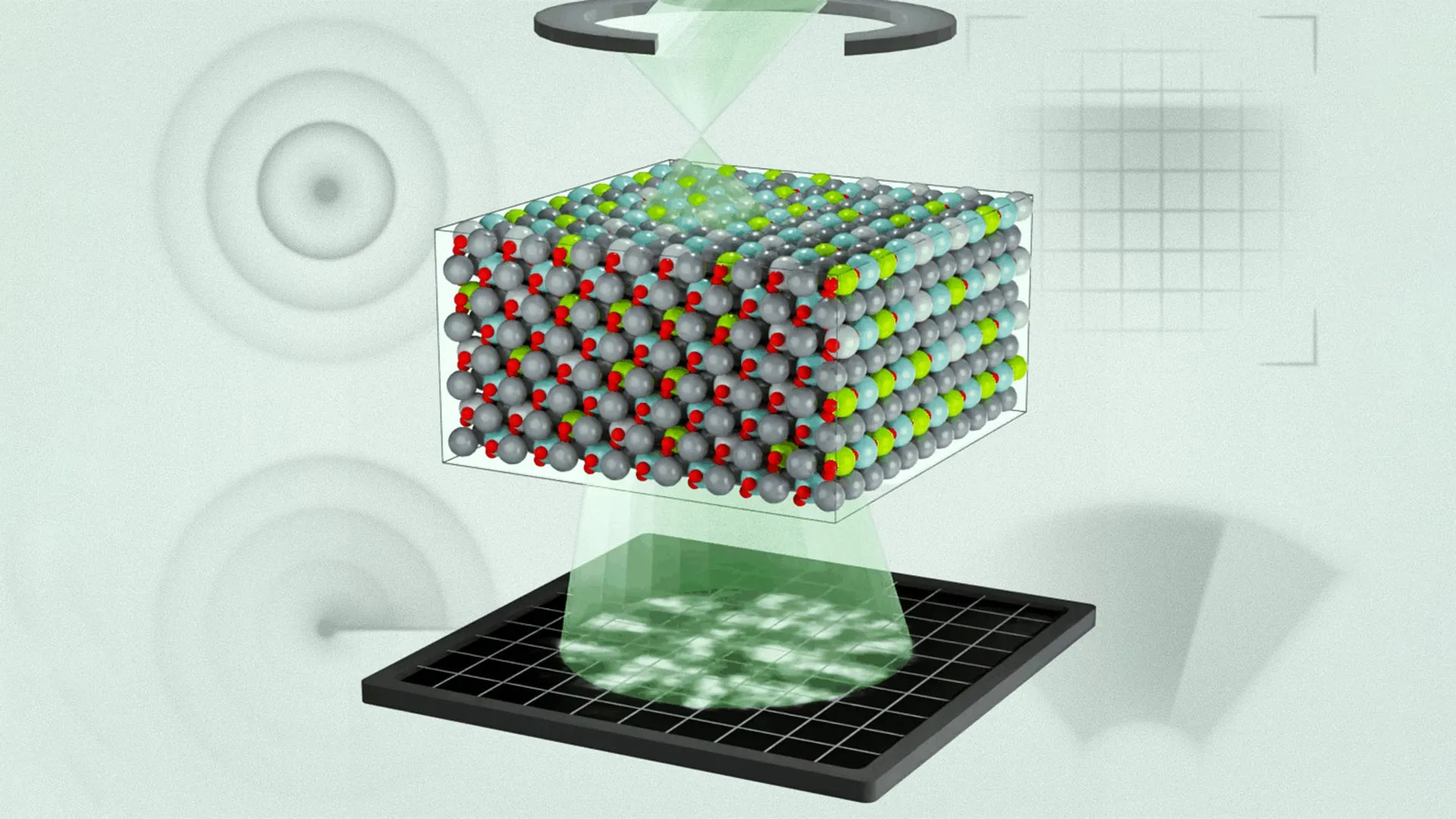
How did the MIT researchers map the 3D structure so precisely, and what techniques did they use?
The team employed a cutting-edge combination of scanning transmission electron microscopy (STEM) and electron ptychography. First, they prepared an ultra-thin specimen of a classic relaxor ferroelectric, lead magnesium niobate-lead titanate (PMN-PT). By scanning a focused electron beam across the sample at atomic resolution, they recorded diffraction patterns from each point. Unlike conventional microscopy that just captures intensity, ptychography uses the interference patterns of scattered electrons to reconstruct both the amplitude and phase of the electron wave. This phase information is crucial because it encodes the electrostatic potential of the atomic columns—effectively, it shows where charges are concentrated. The researchers then collected these 2D slices at different tilts and computationally fused them into a 3D volume with sub-angstrom resolution. This allowed them to visualize individual atoms and the electric fields between them. It was a painstaking process requiring massive data sets and sophisticated algorithms, but the result is the first true 3D nanoscale map of polarization in a relaxor ferroelectric.
What next steps are planned to build on this discovery?
The MIT team's immediate goal is to apply this imaging method to other relaxor compositions, including lead-free alternatives that are safer for the environment. They also want to perform in-situ studies—watching the nanoscale structure change in real time as temperature, electric fields, or mechanical stress are applied. That would reveal the dynamic behavior of polar nanoregions under operating conditions, providing a direct link to device performance. On the theoretical side, the detailed 3D maps are being used to train machine-learning models that can predict new relaxor materials with optimized properties. Collaborators in materials science and electrical engineering are already testing these predictions to synthesize candidate compounds. Ultimately, the hope is to create a 'design toolkit' for relaxor ferroelectrics, where engineers can specify a desired performance parameter (like maximum strain or minimal energy loss) and the toolkit suggests a composition and processing route. This would dramatically shorten the development cycle for next-generation transducers, sensors, and actuators.
Could this breakthrough lead to entirely new types of materials beyond relaxor ferroelectrics?
Absolutely. The 3D mapping technique developed by MIT is not limited to relaxors—it can be applied to any complex material where nanoscale charge ordering is critical. For example, researchers are eyeing multiferroics (materials with simultaneous magnetic and electric ordering) and quantum materials like high-temperature superconductors, where charge stripes and electronic phase separation play a role. The ability to see how dipoles and lattice distortions interact in three dimensions offers a universal window into emergent phenomena. Furthermore, the lessons learned about cooperative polarization in relaxors might inspire synthetic metamaterials—artificial structures that mimic these hierarchical charge patterns. For instance, one could engineer a composite of dielectric nanoparticles embedded in a piezoelectric matrix to create a 'meta-ferroelectric' with tailored properties. In short, this is not just a refinement of an existing material class; it opens a new chapter in how we understand and design materials that harness collective atomic behavior for technology.