Quick Facts
- Category: Science & Space
- Published: 2026-05-05 09:34:38
- How to Leverage Mistral's New Remote Agents and Work Mode in Le Chat
- Navigating AMD's Earnings Surge: A Comprehensive Guide to AI-Driven Growth
- Mythos Preview Shows Leap in Automated Security Exploitation, Researchers Report
- How to Deploy a Fleet of 103 Electric Buses: Lessons from Swedish Cities
- The Art of the Retraction: A Step-by-Step Guide for Ethical Journalism
Introduction
For decades, relaxor ferroelectrics have been the unsung heroes behind medical ultrasound machines, sonar systems, and advanced actuators. Yet, despite their widespread use, the precise arrangement of atoms within these materials remained a frustrating puzzle—until now. In a groundbreaking study, researchers at MIT have successfully mapped the three-dimensional atomic structure of relaxor ferroelectrics in unprecedented detail, revealing hidden patterns in how electric charges organize at the nanoscale. This not only overturns long-held assumptions about the material's behavior but also paves the way for more accurate models to design next-generation electronics. In this step-by-step guide, we'll walk through the process that led to this discovery, from the initial mystery to the final breakthrough.
What You Need (Prerequisites)
Before diving into the steps, it helps to have a basic understanding of key concepts and tools involved in this research. The following are essential for appreciating the journey:
- Relaxor ferroelectrics: A class of materials known for their exceptional piezoelectric and electromechanical properties, but with a complex, disordered atomic structure.
- Advanced imaging techniques: Specifically, electron microscopy and X-ray diffraction methods that can resolve atomic-scale features.
- Computational modeling: Software and algorithms to reconstruct three-dimensional structures from two-dimensional data.
- Basic knowledge of crystallography: Understanding unit cells, lattice parameters, and symmetry.
- Curiosity and patience: Research of this magnitude often involves years of trial and error.
Step-by-Step Process of Discovering the Hidden Structure
Step 1: Identify the Mystery Behind Relaxor Ferroelectrics
The first step in any scientific breakthrough is recognizing what we don’t know. For decades, scientists knew that relaxor ferroelectrics exhibited unusual behaviors, such as a diffuse phase transition and giant piezoelectric responses, but the underlying atomic arrangement was a black box. Traditional crystallography assumed a regular, repeating pattern of atoms, but relaxors defied this—their structure seemed to be disordered yet highly functional. Researchers at MIT began by compiling existing data and pinpointing the key question: how are the electric dipoles arranged at the nanoscale to produce such remarkable properties?
Step 2: Gather Advanced Tools and Techniques
To peel back the layers of this mystery, the team needed more than a standard microscope. They turned to a combination of cutting-edge methods:
- Scanning transmission electron microscopy (STEM): This technique uses a focused electron beam to scan a thin sample, providing atomic-resolution images.
- 3D diffraction mapping: By rotating the sample and collecting diffraction patterns, researchers can reconstruct the reciprocal space.
- Machine learning algorithms: These helped stitch together millions of data points into a coherent three-dimensional model.
Gathering the right tools was crucial—without them, the hidden patterns would remain invisible.
Step 3: Prepare the Sample and Collect Raw Data
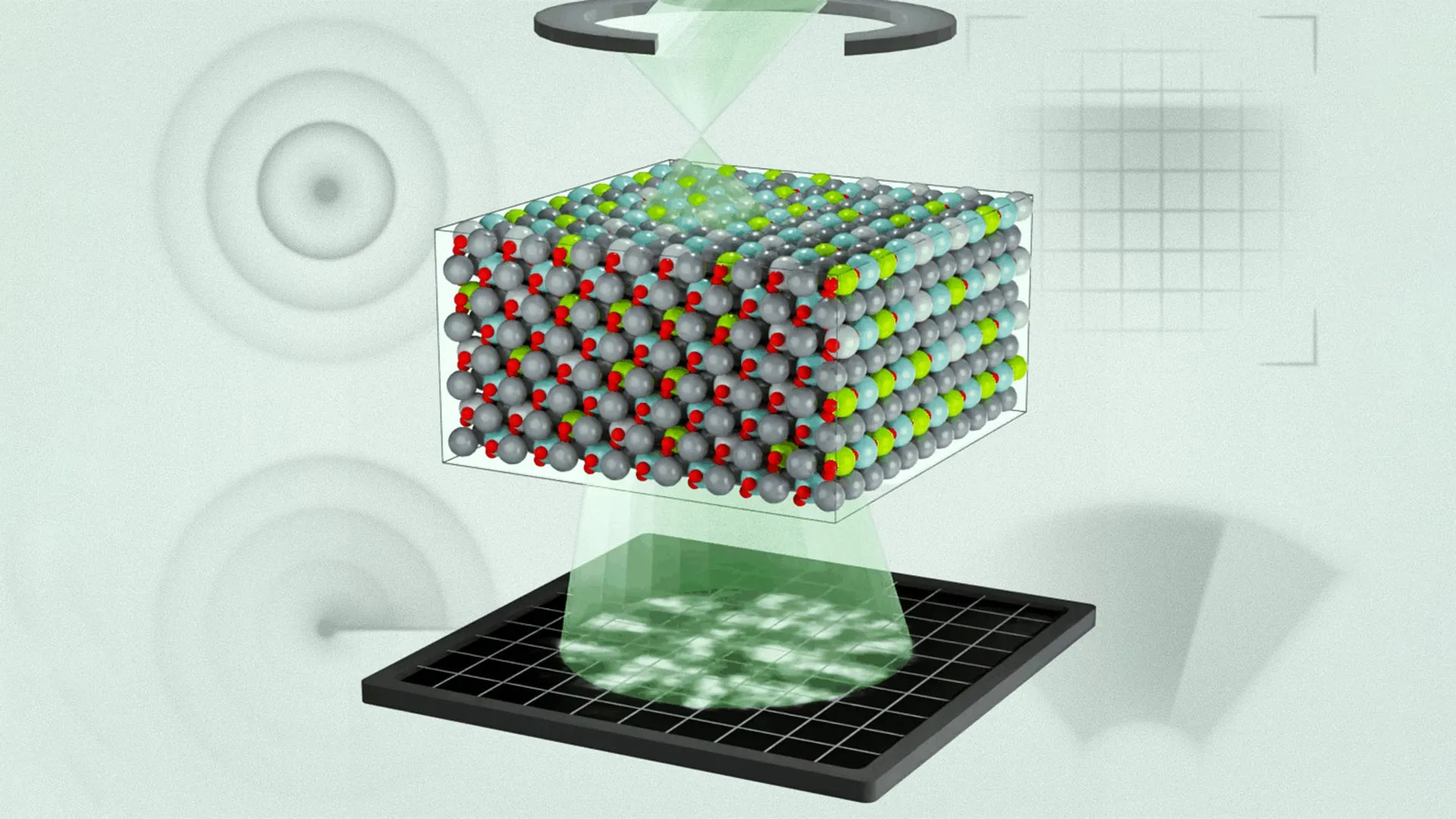
With the tools ready, the next step was to create a pristine sample of a relaxor ferroelectric (commonly lead magnesium niobate-lead titanate, or PMN-PT). The sample had to be ultra-thin (typically less than 100 nanometers thick) to allow electrons to pass through. Using a focused ion beam, scientists sliced a tiny wedge from a single crystal. Then, they mounted it on a special holder and placed it inside the STEM. Over several days, they collected thousands of high-resolution images at various angles, each capturing the positions of individual atoms.
Step 4: Reconstruct the 3D Atomic Arrangement
Raw images are just 2D projections. To see the full structure, the team used a technique called atomic electron tomography. This involves aligning the images from different angles, correcting for distortions, and then using computational algorithms to back-calculate the 3D positions of every atom. The process was computationally intensive—supercomputers ran for weeks to produce a single model. The result was a stunningly detailed map that showed not only where atoms sat but also how they shifted relative to an ideal lattice.
Step 5: Identify Hidden Patterns of Electric Charge
Once the atomic coordinates were known, the next step was to analyze the electric dipole moments—the separation of positive and negative charge centers within each unit cell. By calculating the displacement of cations (like Pb and Ti) from their symmetric positions, the researchers mapped out the nanoscale polarization landscape. To their surprise, they found that the dipoles were not randomly oriented as long assumed. Instead, they formed complex, correlated patterns—like swirls and vortices—that extended over several nanometers. These patterns were the key to the material’s exceptional properties.
Step 6: Challenge Long-Standing Assumptions
The discovery flew in the face of earlier theories, which treated relaxors as having a completely random distribution of dipoles. The hidden order meant that the material’s response to electric fields or mechanical stress was far more cooperative than previously thought. This step involved comparing the new 3D data with predictions from existing models and statistically proving that the patterns were not random. The researchers then published their findings, effectively rewriting the textbooks on relaxor physics.
Step 7: Refine Models for Future Design
Finally, the team used the newly revealed atomic structure to update computational models that predict how relaxors behave under different conditions. With the real 3D arrangement as input, simulations could now accurately reproduce phenomena like the diffuse phase transition and giant piezoelectricity. This step opens the door for engineers to design new relaxor compositions with tailored properties—for example, better sensitivity in ultrasound transducers or higher efficiency in energy-harvesting devices.
Conclusion and Tips
This step-by-step journey from mystery to discovery demonstrates the power of combining advanced experimental techniques with robust computational analysis. For anyone inspired to tackle similar scientific challenges, here are a few tips:
- Embrace interdisciplinary collaboration: The success relied on physicists, materials scientists, and computer scientists working together—don’t isolate yourself.
- Invest in the best tools possible: Sometimes you need to wait for technology to catch up with your questions. Be patient and resourceful.
- Question assumptions: The old theory of random dipoles held for decades because it was convenient. Always test fundamental beliefs with real data.
- Think in 3D: Many properties emerge from the arrangement of atoms in three dimensions—never settle for 2D projections.
- Document every step: Reproducibility is key in science. Keep meticulous records of your methods and data.
With the hidden structure finally revealed, the future of relaxor ferroelectrics looks brighter than ever. Whether you're a student, researcher, or enthusiast, understanding this breakthrough can inspire new ideas in material science, electronics, and beyond.